
OsgoodSchlatter disease and tibial stress fracture Image
Citation, DOI, disclosures and article data. Osgood-Schlatter disease, also known as apophysitis of the tibial tubercle, is a chronic fatigue injury due to repeated microtrauma at the patellar tendon insertion onto the tibial tuberosity, usually affecting boys between ages 10-15 years.

OsgoodSchlatter disease Image
Osgood-Schlatter disease is caused by irritation of the bone growth plate. Bones do not grow in the middle, but at the ends near the joint, in an area called the growth plate. While a child is still growing, these areas of growth are made of cartilage instead of bone. The cartilage is never as strong as the bone, so high levels of stress can.

OsgoodSchlatter disease Image
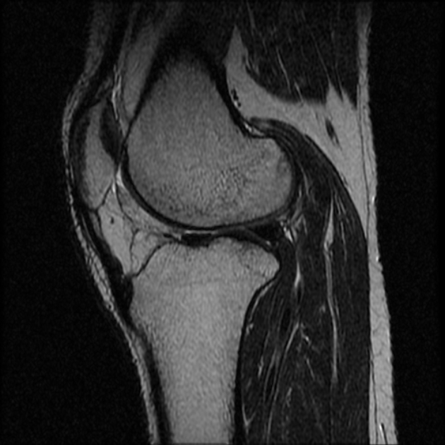
Osgood-Schlatter disease: occurs at the inferior attachment of the patellar tendon onto the tibial tuberosity. Sinding-Larsen-Johansson disease. patellar sleeve fractures: same age group; avulsion of inferior pole cartilage, often with small fracture fragment. infrapatellar bursitis: fluid signal is located anteriorly to the patellar tendon

OsgoodSchlatter disease The Third Eye Radiology site
Case Discussion. Features consistent with Osgood-Schlatter disease. It is a traction apophysitis occurring at the tibial tuberosity because of the pull of the quadriceps muscle group via the patella tendon. It is often seen during rapid growth (8-13 in girls and 12-15 In boys) and is more common in active teenagers.

OsgoodSchlatter disease Image
The Sinding-Larsen Johansson syndrome 1,2 and Osgood Schlatter disorder are osteochondroses of the inferior pole of the patella and the tibial tuberosity respectively. They are associated with activities like running, hiking. They can occur simultaneously as in this case. The main differential diagnosis of Sinding-Larsen Johansson syndrome is.
OsgoodSchlatter's Disease Radiology Qradiology
Purpose: Osgood-Schlatter disease (OSD) is a common cause of anterior knee pain in children. The natural history of childhood OSD has been described in detail in the literature, and it has been shown to be a largely self-limiting condition which resolves with closure of the growth plates. However, some children with OSD continue to suffer with anterior knee pain into adulthood.

OsgoodSchlatter disease Image
About Radiopaedia.org. Radiopaedia.org is a rapidly growing, peer-reviewed open-edit radiology resource, compiled by radiologists and other health professionals from across the globe. Radiopaedia's mission is to create the best radiology reference the world has ever seen and to make it available for free, forever, for all.
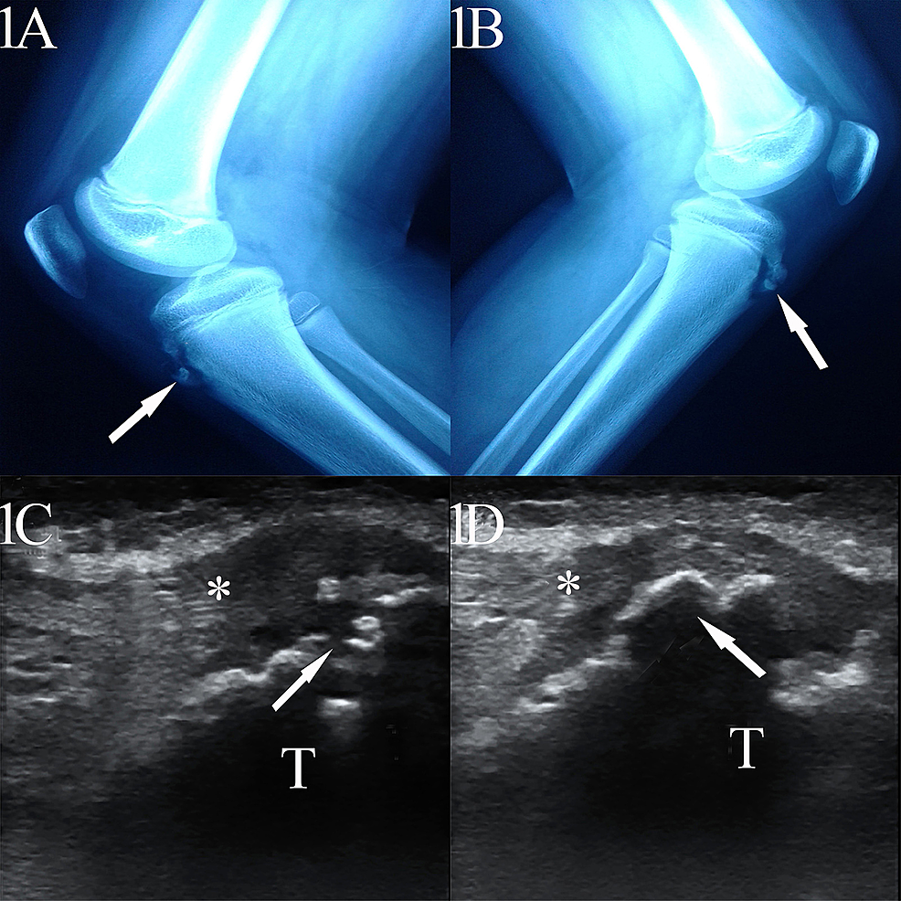
Cureus OsgoodSchlatter Disease Unveiled Under Highfrequency
Gender: Male. x-ray. Lateral. Frontal. Bone fragmentation at the tibial tubercle with soft tissue swelling keeping with Osgood-Schlatter disease. Annotated image.

Image
Osgood-Schlatter disease (OSD), also known as Lannelongue disease [ 1 ], is a type of osteochondrosis first described by Osgood and Schlatter in 1903 [ 2 ]. It consists of the onset of a traction apophysitis as a consequence of repeated contractions of the femoral rectum part of the quadriceps [ 3] (see Figure 1) and may be bilateral [ 4 ]. OSD.

OsgoodSchlatter disease Radiology Case
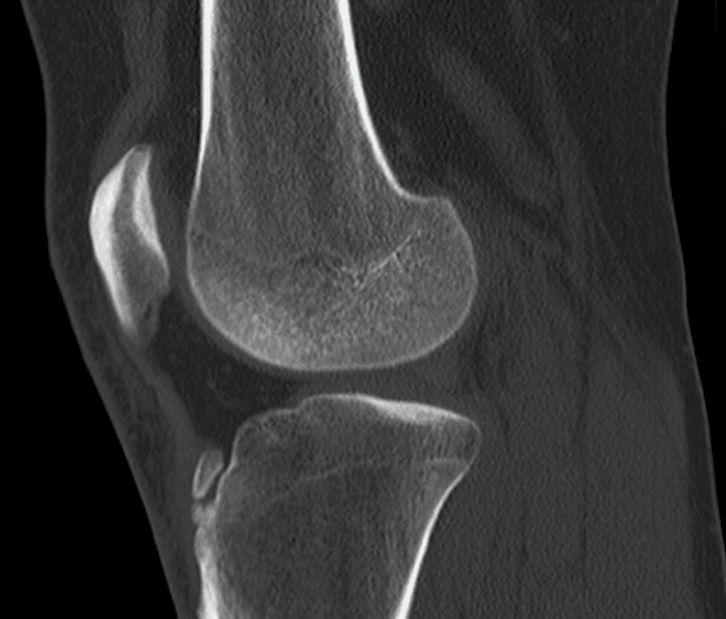
Fragmented apophysis at tibial tuberosity. Overlying soft tissue swelling is noted. Soft tissue haziness is noted in inferior most part of fat pad; obliterating shadow of inferior most part of patellar ligament.

OsgoodSchlatter disease Radiology Case
Osgood-Schlatter disease. Osgood-Schlatter disease, also known as apophysitis of the tibial tubercle, is a chronic fatigue injury due to repeated microtrauma at the patellar tendon insertion onto the tibial tuberosity, usually affecting boys between ages 10-15 years. Terminology Unresolved Osgood-Schlatter disease is t.

OsgoodSchlatter disease Image
Case Discussion. Features on X-ray in 10 years patient with knee pain consistent with Osgood-Schlatter disease , a chronic fatigue injury due to repeated microtrauma at the patellar ligament insertion onto the tibial tuberosity.

Osgood Schlatter disease Image
A lateral view of the right knee was obtained as a comparison with the left, which demonstrated a fragmented tibial apophysis which is not diagnostic for Osgood Schlatter disease. The non-ossifying fibroma on the right side was an incidental finding. In this case, the fragmented apophysis is in keeping with the clinical symptoms of Osgood.
OsgoodSchlatter disease Image
Free Shipping on eBay

OsgoodSchlatter disease Image
Osgood Schlatter disease, also known as osteochondrosis, tibial tubercle apophysitis, or traction apophysitis of the tibial tubercle, is a common cause of anterior knee pain in the skeletally immature athletic population. Clinical presentation classically associates atraumatic, insidious onset of anterior knee pain, with tenderness.

OsgoodSchlatter disease Image
SINCE Osgood described a lesion of the adolescent tibial tuberosity in 1903 (1) various men have collected small series of cases and each has given his opinion as to the etiology. Again, Schlatter, in 1908 (2), described a similar condition which is now recognized as a clinical entity and generally known as "Osgood-Schlatter's disease," though occasionally as "Schlatter's disease." It.
